oct b scan
The optic nerve head is also visible at the back of your eye so an OCT scan can also evaluate any disorders of the optic nerve. OCT B-scan of the blood circulation Each group of OCT B-scan generates an image of the blood flow.

Polypoidal Choroidal Vasculopathy Pcv Nursing Flashcards Optician Training Eye Health
From Wikimedia Commons the free media repository.

. 19 The A-scan ultrasonogram usually demonstrates moderate to high internal reflectivity. File usage on other wikis. FileOCT B-Scan Setup-ensvg.
16 17 CHAPTER 2 CHOROIDAL NEOVASCULARIZATION OCT-Angiography is an extraordinary tool for visualizing abnormal choroidal vasculature. 275 240 pixels 551 480 pixels 881 768 pixels 1175 1024 pixels 2349 2048 pixels. An optical coherence tomography scan commonly referred to as an OCT scan helps us to view the health of your eyes in greater detail by allowing us to see whats going on beneath the surface of the eye.
The OCT b-scan demonstrates a retinal pigment epithelial detachment RPED subretinal fluid an intraretinal cyst and hyper-reflective material characteristic of CNV. Comparison to the patients pre- and post-treatment OCT B-scans showed that there was a characteristic sign of PCV. To describe a novel imaging technique which we call dense B-scan optical coherence tomography angiography DB OCTA in which thin dense raster scans are used to produce highly resolved structural B-scans with superimposed flow signal that provide precise correlation between retinal microstructure and blood flow.
The resolution obtained by OCT is 1-2 orders of magnitude higher than ultrasound and has been demonstrated to work on both transparent and non-transparent tissue with high reflection. Overlying retinal detachment is common and sound attenuation in the lesion is usually moderate13 In unusual cases choroidal detachment may be seen. Size of this PNG preview of this SVG file.
An OCT scan can help your optician to see whats going on beneath the surface of your eye providing a picture of the layers of your retina. B-OCT measures ocular axial dimensions in four 3 mm wide and 25 mm deep measurement windows. In OCT many one-dimensional scans a-scans are performed at several depths to create a two-dimensional image b-scan.
The B-scan ultrasonogram shows an echogenic subretinal mass with diffuse ill-defined borders. OCT-Angiography map These maps are a reconstruction of the microvasculature of the retina and choroid. Imagine it like a cake we can see the top of the cake and the icing using the 2D digital retinal photography fundus camera but the.
An inverted U-shaped elevation of the retinal pigment epithelium RPE that was usually located between the RPE and Bruch membrane. Your optician can then map out the layers and measure the thickness. The location of this feature on OCT B-scans correlated to the sites of polypoidal lesions on ICGA.
A4-6 Fluorescein angiography FA early intermediate and late frames showing increasing hyper-fluorescence and staining of the CNV. In contrast B-scans for conventional OCT are derived from sagittal and transverse sections. 687 599 pixels.
The use of OCT B-scan imaging can facilitate more widespread diagnosis of PCV and can significantly affect the management approach to include the choice of anti-VEGF agent or the initiation of combination therapy with PDT and intravitreal anti-VEGF injections which may lead to potential visual benefits and decreased treatment for PCV eyes. File usage on Commons. OCT B scan of a normal eye imaged using SS-OCT.
In the upper and central parts of the figure a single horizontal and vertical scan of ocular structures in their respective measurement windows is shown. The B scan is 16 mm in length and offers within a single frame clear visualization of the vitreous retina choroid and. The use of en-face OCT for ARPE allows a frontal layer-by-layer visualization of the retina.
The lower row presents the corresponding A-scan. The advantage of this type of presentation B-Scan is that both the length of the flaw and its depth below the surface is revealed. En-face OCT is a novel imaging technique generating frontal scans derived from SD-OCT 4.
B- Scan Display The B-scan display as shown in the above figure right-hand side shows reflection cross-sectional view from the top and bottom of the test object flaws as the probe moves along a line one axis.
Pin On Patient Information Pages

Ophthalmic B Scan Optometry New Things To Learn Eyes

The Anatomy Of An Oct Scan Optometry Education Optometry Anatomy

Mesh Stigmergy

Ophthalmology Management Cirrus Hd Oct Today And Tomorrow

The Official Oct Interpretation Optometry School Optometry Education Opthalmic Technician

Figure 1 Vascular Probe Continuity

Pin On Patient Information Pages

Mudher Abdulla Adli Kullanicinin Optical Coherence Tomography Oct Panosundaki Pin

Macula Eye Anatomy Eye Facts Macular Degeneration Facts

Macular Pucker Numerous Underlying Conditions Lead To Scar Tissue On The Macula When This Affects Vision The Scarring Can B Scar Tissue Pathology Restoration

Figure 1 The Triangular Subretinal Hyporeflective Space In Papilledema A B And The Buried Optic Nerve Head Drusen In Pse Study Guide Optic Nerve View Image
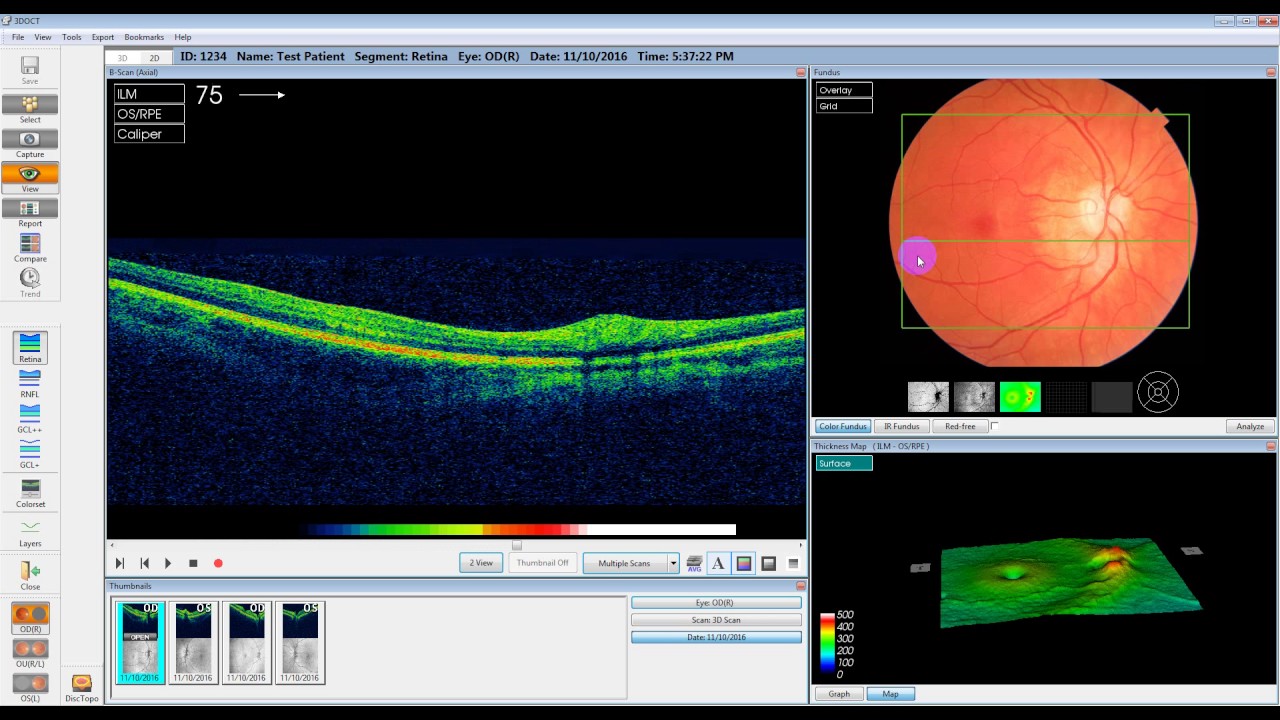
Maestro Software Tutorial

Pin Page

Pin On Casa Aperta Daybed

Pin On Glaucoma

Bhavin Jankharia On Instagram Mars Ct Or Mri This Patient Has Come 10 Years Post Elbow Replacement With A Growing Volar Swellin Radiology Imaging Pet Ct Mri

Demonstration Of Choroidal Neovascularization Associated With An Intraretinal Lesion On Indocyanine Green Ang Optical Coherence Tomography Eye Health Optometry

Pin On Ophthalmology


Comments
Post a Comment